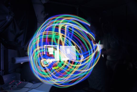
About 100 years ago, humanity learned to see with the help of electrons. In 1924, Louis de Broglie posited that – like light particles – electrons have wave properties. In 1927, the US physicists Davisson and Germer provided experimental proof of this. A few years later, the engineers Ernst Ruska and Max Knoll built the first electron microscope, which was more powerful than any light microscope. Given that electron waves are diffracted by much smaller objects than photons, the optical resolution limit of light was surmounted, heralding a new era of microscopy.
Combining two worlds: Quantum electron microscopy
“Electron microscopy is a crazy, cool technique,” Philipp Haslinger, Associate Professor at the TU Wien, says enthusiastically. “In principle, we could use it to look at the spike proteins of a virus or its DNA – at the level of atoms, the pixels of reality.” Haslinger, a quantum optics specialist, deliberately says “could”, because there is a catch: the electrons are typically so high in energy that they destroy sensitive samples. For this reason, biological processes cannot be observed “live” with electron microscopes.
According to Haslinger, there is one possible solution: “Gaining more information from fewer electrons.” In pursuit of this goal, his eleven member team uses “quantum electron microscopy”, which combines classic electron microscopy with the newer world of photon-based quantum optics.
Spooky imaging
One of their possible ideas is based on a method going by the evocative name of “quantum ghost imaging” or Zou-Wang-Mandel, opens an external URL in a new window. In this method, an entangled electron-photon pair generates the image of the object. This is how it works: first, an electron races through a translucent medium and “overtakes” the light there, “a bit like an airplane going supersonic,” explains Haslinger. This creates a photon which is taken to be entangled with the electron. While the electron travels towards the sample, the photon enters a camera detector. As the two are entangled, the photon can be used to measure whether the electron has hit the sample. If the detected photons can be space-resolved successfully, the image of the object can be constructed.
At least, this is the theory behind the approach. “Several research groups around the world are working on establishing the first proof of this entanglement – and we are up in the front line,” says Haslinger. In practice, the innovative ideas are fraught with technical challenges. The team first had to adjust the existing microscope. “Normally, electron microscopes are built completely sealed from light – but we drill holes in them so that photons can escape so as to be measured,” smiles the physicist.
Promising outlook for biology and materials science
What is needed now is proof of principlethat the method can generate electron-photon pairs. “In fact, it could happen any day now,” hopes Haslinger. “We have already recorded a ghost image. So we were able to see with electrons what the photon 'saw'. Now we are looking for evidence of interference phenomena between the two particles. Finding this evidence would give us clear smoking-gun proof of entanglement.”
An established variant of ghost imaging that uses entangled photon-photon pairs has proven its worth when observing particularly light-sensitive objects. If Haslinger's plan works out, this sparing treatment of the sample could for the first time be combined with the high optical resolution of electrons. Such a development would open up promising applications, for example in battery research: the molecular and atomic changes on the surfaces of materials during charging and discharging could be better observed and this would help to identify optimized materials. There might also be spectacular new insights in biology, such as observing proteins as they fold without their being broken during irradiation. “Watching life as it happens, that would be a dream,” beams Haslinger. A good twenty years ago, as a young physics student, he attended lectures by Anton Zeilinger, who got him interested in quantum optics. Now he and his colleagues could bring a new quality to electron microscopy, the history of which began a century ago.
Personal details
Philipp Haslinger, opens an external URL in a new window is Associate Professor for Atomic Interferometry at the TU Wien. His research group focuses on the development of novel quantum tools for matter wave optics with atoms and electrons and conducts research on various dark energy models. Haslinger completed his PhD at the TU Wien in 2013. He then spent time at UC Berkeley on an FWF fellowship before returning to Vienna to continue his research at the Atomic Institute of TU Wien. The project “Quantum optics with electron-photon pairs” is funded as a stand-alone project by the FWF. In 2018, he was awarded the FWF START Prize, opens an external URL in a new window. He is involved in outreach projects, opens an external URL in a new window at the interface between art and science.
Publication
Philipp Haslinger et al.: Spin resonance spectroscopy with an electron microscope, opens an external URL in a new window, in Quantum Science and Technology 2024
Contact
Prof. Philipp Haslinger
TU Wien
Institute of Atomic and Subatomic Physics
+43 1 58801 141868
philipp.haslinger@tuwien.ac.at , opens an external URL in a new window
Text: Ingrid Ladner, Austrian Science Fund FWF
